Mapping the Brain on a Massive Scale
September 28, 2010 | Source: Technology Review

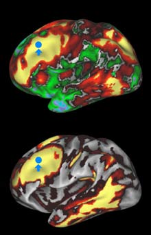
In the top image, areas in yellow and red are structurally connected to the area indicated by the blue spot. In the bottom image, areas in yellow and red are those that are functionally connected to the blue spot. (David Van Essen, Washington University)
The National Institutes of Health announced $40 million in funding this month for the five-year effort, dubbed the Human Connectome Project. Scientists will use new imaging technologies, some still under development, to create both structural and functional maps of the human brain.
The researchers also plan to collect genetic and behavioral data, testing participants’ sensory and motor skills, memory, and other cognitive functions, and deposit this information along with brain scans in a public database that can be used to search for the genetic and environmental factors that influence the structure of the brain.
David Van Essen, a neuroscientist at Washington University and his collaborators plan to scan participants using two relatively recent variations on MRI. Diffusion imaging, which detects the flow of water molecules down insulated neural wires, indirectly measures the location and direction of the fibers that connect one part of the brain to another. Functional connectivity, in contrast, examines whether activity in different parts of the brain fluctuates in synchrony. The regions that are highly correlated are most likely to be connected, either directly or indirectly. Combining both approaches will give scientists a clearer picture.
