Integrating biological components into electronic circuits
June 9, 2011
Researchers at the University of Pennsylvania have developed a way to form biological molecules that can be directly integrated into electronic circuits, and a microscope technique to measure them.
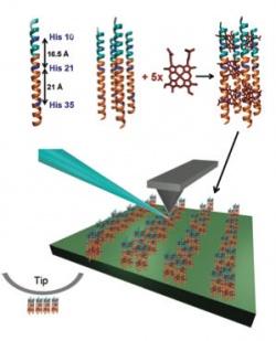
The development involves artificial proteins, bundles of peptide helices with a photoactive molecule inside. These proteins are arranged on electrodes, a common feature of circuits that transmit electrical charges between metallic and non-metallic elements. When light is shined on the proteins, they convert photons into electrons and pass them to the electrode.
The researchers also developed a new kind of atomic force microscope technique, known as torsional resonance nanoimpedance microscopy. They used a metallic tip and put an oscillating electric field on it. By seeing how electrons react to the field, they were able to measure more complex interactions and more complex properties, such as capacitance.
They stamped the self-assembling proteins onto sheets of graphite electrodes. This manufacturing principle and the ability to measure the resulting devices could have a variety of applications including photovoltaics, the researchers said.
Ref.: Kendra Kathan-Galipeau, et al., Direct Probe of Molecular Polarization inDe NovoProtein-Electrode Interfaces, ACS Nano, 2011; 110603081000090 [DOI: 10.1021/nn200887n]

