Nanopatterned natural biological scaffold for stem cells may allow for softer engineered tissues
February 26, 2014

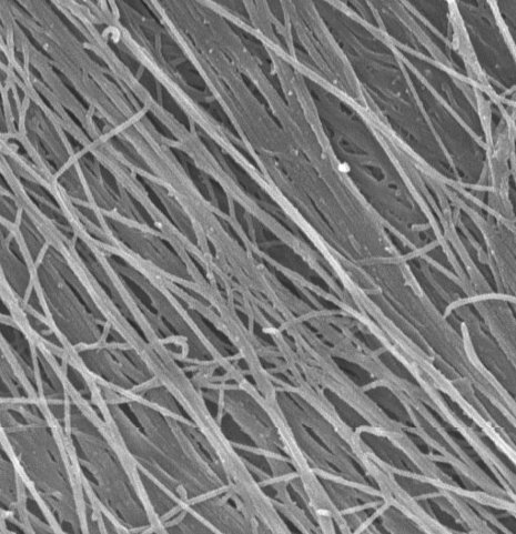
Highly aligned nanofibers created by fibroblasts form a biological scaffold that could prove an ideal foundation for engineered tissues. Stem cells placed on the scaffold thrived, and it had the added advantage of provoking a very low immune response. (Credit: MTU)
Feng Zhao of Michigan Technological University (MTU) has persuaded fibroblasts — cells that make up the extracellular matrix in the body — to make a well-organized nanopatterned scaffold (support structure). This discovery could have major implications for growing engineered tissue to repair or replace virtually any part of our bodies.
In all multicellular organisms, including people, cells make their own extracellular matrix, a complex, nurturing structure that is essential for many biological functions, including growth and healing. But in the lab, scientists attempting to grow tissue must provide an artificial scaffold.
Typically, researchers construct scaffolds from synthetic materials or natural animal or human substances grown in a Petri dish. (KurzweilAI has reported a variety of such synthetic approaches to creating scaffolds for extracellular matrices.) But such scaffolds have not been able to mimic the highly organized structure of the matrix made by living things — at least until now.
Engineering softer tissues
Zhao’s fibers are a mere 80 nanometers across, similar to fibers in a natural biological matrix. And, since her scaffold is made by cells, it is composed of the same intricate mix of all-natural proteins and sugars found in the body. Plus, its nanofibers are as highly aligned as freshly combed hair.
The trick was to orient the cells on a nano-grate (130 nm in depth) that guided their growth and the creation of the scaffold. “The cells did the work,” Zhao said. “The material they made is quite uniform, and of course it is completely biological.”
Stem cells placed on her scaffold thrived, and it had the added advantage of provoking a very low immune response.
“We think this has great potential,” she said. “I think we could use this to engineer softer tissues, like skin, blood vessels and muscle.”
The work is described in the paper “Highly Aligned Nanofibrous Scaffold Derived from Decellularized Human Fibroblasts,” coauthored by Zhao, postdoctoral researcher Qi Xing and undergraduate Caleb Vogt of MTU and Kam W. Leong of Duke University and published Jan. 29 in Advanced Funcational Materials. Zhao designed the project. Xing and Vogt did the work, and Leong developed the template for cell growth.
Other Duke University researchers took a different approach, combining a synthetic scaffolding material with gene delivery techniques to direct stem cells into becoming new cartilage, as KurzweilAI reported on Feb. 21. Could gene delivery enhance the MTU process?
The work was funded by the National Institutes of Health, the Flight Attendant Medical Research Institute, and Michigan Technological University.
Abstract of Advanced Functional Materials paper
Native tissues are endowed with a highly organized nanofibrous extracellular matrix (ECM) that directs cellular distribution and function. The objective of this study is to create a purely natural, uniform, and highly aligned nanofibrous ECM scaffold for potential tissue engineering applications. Synthetic nanogratings (130 nm in depth) are used to direct the growth of human dermal fibroblasts for up to 8 weeks, resulting in a uniform 70 μm-thick fibroblast cell sheet with highly aligned cells and ECM nanofibers. A natural ECM scaffold with uniformly aligned nanofibers of 78 ± 9 nm in diameter is generated after removing the cellular components from the fibroblast sheet. The elastic modulus of the scaffold is well maintained after the decellularization process because of the preservation of elastin fibers. Reseeding human mesenchymal stem cells (hMSCs) shows the excellent capacity of the scaffold in directing and supporting cell alignment and proliferation along the underlying fibers. The scaffold’s biocompatibility is further examined by an in vitro inflammation assay with seeded macrophages. The aligned ECM scaffold induces a significantly lower immune response compared to its unaligned counterpart, as detected by the pro-inflammatory cytokines secreted from macrophages. The aligned nanofibrous ECM scaffold holds great potential in engineering organized tissues.
