New sensors monitor brain activity and blood flow deeper in the brain with high sensitivity and high speed
April 30, 2018

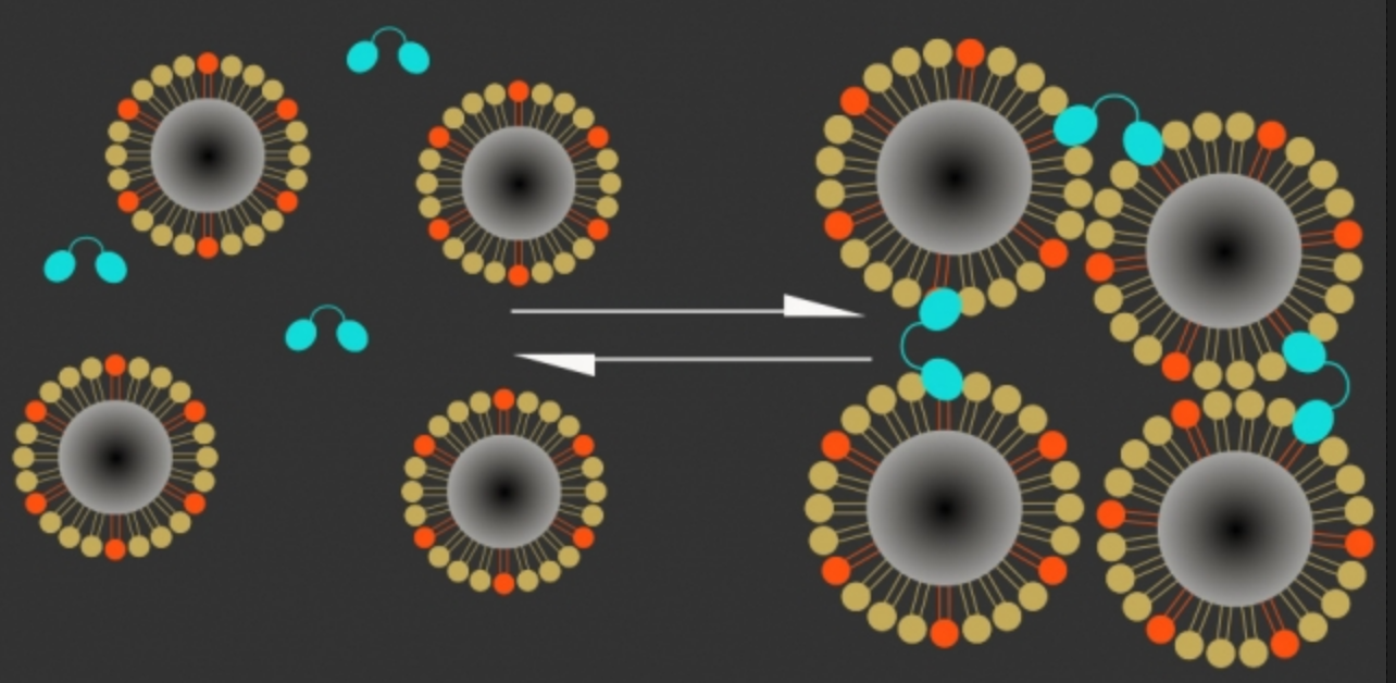
Magnetic calcium-responsive nanoparticles (dark centers are magnetic cores) respond within seconds to calcium ion changes by clustering (Ca+ ions, right) or expanding (Ca- ions, left), creating a magnetic contrast change that can be detected with MRI, indicating brain activation. (High levels of calcium outside the neurons correlate with low neuron activity; when calcium concentrations drop, it means neurons in that area are firing electrical impulses.) Blue: C2AB “molecular glue” (credit: The researchers)
Calcium-based MRI sensor enables deep brain imaging
MIT neuroscientists have developed a new magnetic resonance imaging (MRI) sensor that allows them to monitor neural activity deep within the brain by tracking calcium ions.
Calcium ions are directly linked to neuronal firing at high resolution — unlike the changes in blood flow detected by functional MRI (fMRI), which provide only an indirect indication of neural activity. The new sensor can also monitor large areas, compared to fluorescent molecules, used to label calcium in the brain and image it with traditional microscopy, which is limited to small areas of the brain.
A calcium-based MRI sensor could allow researchers to link specific brain functions directly to specific neuron activity, and to determine how distant brain regions communicate with each other during particular tasks. The research is described in a paper in the April 30 issue of Nature Nanotechnology. Source: MIT

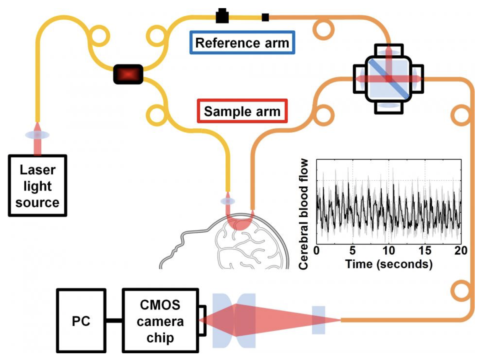
New technique for measuring blood flow in the brain uses laser light shined into the head (“sample arm” path) through the skull. The return signal is boosted by a reference light beam and returned to a detector camera chip. (credit: Srinivasan lab, UC Davis)
Measuring deep-tissue blood flow at high speed
Biomedical engineers at the University of California, Davis, have developed a more-effective, lower-cost technique for measuring deep tissue blood flow in the brain at high speed. It could be especially useful for patients with stroke or traumatic brain injury.
The technique, called “interferometric diffusing wave spectroscopy” (iDWS), replaces about 20 photon-counting detectors in diffusing wave spectroscopy (DWS) devices (which cost a few thousand dollars each) with a single low-cost CMOS-based digital-camera chip.
The NIH-funded work is described in an open-access paper published April 26 in the journal Optica. Source: UC Davis
