New technique takes a big step in examination of small structures
March 6, 2008 | Source: KurzweilAI
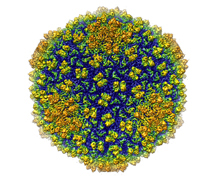
Researchers from Purdue University, Baylor College of Medicine, and MIT captured a three-dimensional image of a live virus at a resolution of 4.5 angstroms, tracing for the first time the polypeptide chain structure of a live virus.

bacteriophage Epsilon15
The technique used, single-particle electron cryomicroscopy, maintains the sample in a natural state. X-ray crystallography, for example, requires the sample to be crystallized.
